Anatomy Of Musckes Sndctendons - Human arm muscle anatomy in detail : It is comprised of two bones:. The fleshy, thick part of the muscle is called its belly. The muscles you probably know the best are your glutes. It is separated from the first layer of muscles by the lateral plantar vessels and nerve. The tendons for these muscles begin at your ischial tuberosity, or ischium (the bony bump under each buttock), and attach on the outer edges of your shinbones (tibia and fibula) just below the back of your knee. The smaller bone that runs alongside the tibia (fibula) and the.
The peroneal muscles (peroneus longus and peroneus brevis), on the outside edge of the ankle and foot. Four muscles and their attached tendons make up the rotator cuff. The knee joint is most significantly affected by two major muscle groups: The majority of muscles in the leg are considered long muscles, in that they stretch great distances. There are tendons in your elbow that attach muscle to bone.
Anatomy Of The Coracobrachialis Muscle ... from orthopaedicprinciples.com The tendons are the attachment of the muscle to the bone. *the origin, insertion, and belly.* a muscle's origin is where a tendon attaches it to the *less* movable bone. Foot_anatomy_muscles_and_tendons 2/14 foot anatomy muscles and tendons encyclopedia of human anatomic variation is the long awaited update to this classic reference. The wrist joint is a complex joint which connects the forearm to the hand, allowing a wide range of movement. Every skeletal muscle has three main parts: Lesson on the anatomy of the forearm: A tendon connects the muscle to the bone. The muscles you probably know the best are your glutes.
The wrist joint is a complex joint which connects the forearm to the hand, allowing a wide range of movement.
They are the continuations of muscles and allow them to connect to bones. Each of them aids in a specific motion of your shoulder. The quadriceps muscles provide strength and power with knee extension (straightening). The posterior upper leg muscles provide your knees with mobility (extension, flexion and rotation) and strength. Lesson on the anatomy of the forearm: Maybe you would like to learn more about one of these? The knee is one of the largest and most complex joints in the body. The shoulder is not a single joint, but a complex arrangement of bones, ligaments, muscles, and tendons that is better called the shoulder girdle. Nerves and blood vessels that supply the bones and muscles of the hip. The knee joint is most significantly affected by two major muscle groups: Early 2008 this issue of foot and ankle clinics is dedicated to the cavovarus. On the other hand, the insertion is where a tendon attaches that muscle to the *more* movable bone. The thigh bone or femur and the pelvis which is made up of three bones called ilium, ischium, and pubis.
The gastrocnemius and soleus muscles taper and merge at the base of the calf muscle. The quadriceps muscles provide strength and power with knee extension (straightening). The knee is one of the largest and most complex joints in the body. Every skeletal muscle has three main parts: As these muscles contract and relax, they move skeletal bones to create movement of the body.
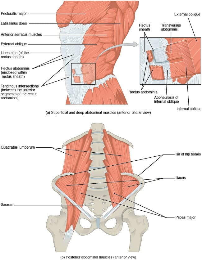
Abdomen (Anatomy): Definition, Function, Muscles | Biology ... from biologydictionary.net Nerves and blood vessels that supply the bones and muscles of the hip. The shoulder girdle includes three bones—the scapula, clavicle and humerus. The primary function of the shoulder girdle is to give strength and range of motion to the arm. The quadriceps muscles provide strength and power with knee extension (straightening). There are numerous tendons around the knee that also help to stabilize the knee. When the muscle contracts, the tendons are pulled, and the bone is moved. In this lesson, we look at the muscle. Every skeletal muscle has three main parts:
There are numerous tendons around the knee that also help to stabilize the knee.
Although the majority of the muscle mass is located anteriorly to the humerus, it has no attachment to the bone itself. The important tendons of the elbow are the biceps tendon, which is attached the biceps muscle on the front of your arm, and the triceps tendon, which attaches the triceps muscle on the back of your arm. The hip joint is the junction where the hip joins the leg to the trunk of the body. The muscles of the abdomen, lower back, and pelvis are separated from those of the chest by the muscular wall of the diaphragm, the critical breathing muscle. There are a number of tendons located in the foot and ankle all responsible for different ankle, foot and toe movements. Major muscles of the ankle. The quad muscles— which form the meaty mass on the front of your thighs — are among your strongest muscle groups, and play a critical role in athletic activities. The quadriceps muscles provide strength and power with knee extension (straightening). In this lesson, we look at the muscle. There are tendons in your elbow that attach muscle to bone. The muscles you probably know the best are your glutes. The shoulder girdle includes three bones—the scapula, clavicle and humerus. All together they help hold your upper arm in place in the shoulder.
Lesson on the anatomy of the forearm: This is lesson 1 on the anatomy of the forearm. It is comprised of two bones: There are a number of tendons located in the foot and ankle all responsible for different ankle, foot and toe movements. The muscles you probably know the best are your glutes.
Pin on School- PTA from i.pinimg.com Tendons are elastic tissues made up of collagen. The quad muscles— which form the meaty mass on the front of your thighs — are among your strongest muscle groups, and play a critical role in athletic activities. The peroneal muscles (peroneus longus and peroneus brevis), on the outside edge of the ankle and foot. It is comprised of two bones: On the other hand, the insertion is where a tendon attaches that muscle to the *more* movable bone. All together they help hold your upper arm in place in the shoulder. Leg extensors, posterior leg muscles, extrinsic foot muscles, and intrinsic foot muscles. Nerves and blood vessels that supply the bones and muscles of the hip.
Early 2008 this issue of foot and ankle clinics is dedicated to the cavovarus.
Tendons also help to provide stability around the foot and ankle Foot_anatomy_muscles_and_tendons 2/14 foot anatomy muscles and tendons encyclopedia of human anatomic variation is the long awaited update to this classic reference. The thigh bone or femur and the pelvis which is made up of three bones called ilium, ischium, and pubis. The peroneal muscles (peroneus longus and peroneus brevis), on the outside edge of the ankle and foot. Lying exposed between the protective bones of the superiorly located ribs and the inferiorly located pelvic girdle, the muscles of this region play a critical role in protecting the. Originates from the medial and lateral plantar surface of the calcaneus. The knee joint is most significantly affected by two major muscle groups: The knee joins the thigh bone (femur) to the shin bone (tibia). The quadratus plantae muscle is located superior to the flexor digitorum longus tendons. The gastrocnemius and soleus muscles taper and merge at the base of the calf muscle. Lesson on the anatomy of the forearm: The posterior upper leg muscles provide your knees with mobility (extension, flexion and rotation) and strength. All together they help hold your upper arm in place in the shoulder.